
Shoulder Arthroscopy - CAM 774
Description
General Information
- It is an expectation that all patients receive care/services from a licensed clinician. All appropriate supporting documentation, including recent pertinent office visit notes, laboratory data, and results of any special testing must be provided. If applicable: All prior relevant imaging results and the reason that alternative imaging cannot be performed must be included in the documentation submitted.
- The guideline criteria in the following sections were developed utilizing evidence-based and peer-reviewed resources from medical publications and societal organization guidelines as well as from widely accepted standard of care, best practice recommendations.
Purpose
This guideline addresses elective, non-emergent, arthroscopic shoulder repair procedures, including Rotator Cuff Repair, Labral Repairs, Lysis of Adhesions (Capsulotomy), Distal Clavicle Excision (DCE), Long Head Biceps (LHB) Tenotomy or Tenodesis, Loose body removal, Synovectomy, and Subacromial Decompression (SAD).
Scope
Surgical indications are based on relevant subjective clinical symptoms, objective physical exam & radiologic findings, and response to previous non-operative treatments when medically appropriate.
Open, non-arthroplasty shoulder repair surgeries are performed as dictated by the type and severity of injury and/or disease.
In order for surgeries to be considered medically necessary there must be clear medical records that demonstrate a clear surgical plan that matches the request for surgery.
GENERAL REQUIREMENTS
Elective surgery of the shoulder may be considered if the following general criteria are met:
- Clinical correlation of individual’s subjective complaints with objective exam findings and/or imaging (when applicable)
- Individual has limited function (age-appropriate activities of daily living (ADLs), occupational, or athletic)
- Individual is medically stable and optimized for surgery, and any treatable comorbidities are adequately medically managed such as diabetes, nicotine addiction, or an excessively high BMI. There should also be a shared decision between the patient and physician to proceed with shoulder surgery when comorbidities exist as it pertains to the increased risk of complications.
- Individual does not have an active local or systemic infection
- Individual does not have active, untreated drug dependency (including but not limited to narcotics, opioids, muscle relaxants) unless engaged in a treatment program
- A smoking cessation program is highly recommended for all actively smoking patients (1,2)
Clinical notes should address:
- Symptom onset, duration, and severity
- Loss of function and/or limitations
- Type and duration of non-operative management modalities (where applicable)
Non-operative management, when required, will be specified within the clinical indications below and may include one or more of the following:
- Physical therapy or properly instructed home exercise program
- Rest or activity modification
- Application of heat or ice
- Minimum of 4 weeks of oral NSAIDs (if not medically contraindicated)
- Single injection of corticosteroid and local anesthetic into subacromial, intra-articular space, or bicipital groove
Policy
INDICATIONS
Diagnostic Shoulder Arthroscopy
Diagnostic arthroscopy is considered medically necessary when the following criteria in either section have been met (3,4):
- Section One
- For the evaluation of a painful total shoulder arthroplasty
- Section Two
- Severe, disabling pain and/or a documented loss of shoulder function which interferes with the ability to carry out age-appropriate activities of daily living and/or demands of employment
- Individual demonstrates any of the following abnormal, shoulder physical examination findings, as compared to the non-involved side:
- Functionally limited range of motion (active or passive)
- Measurable loss in strength
- Positive impingement signs
- Individual has undergone an appropriate radiographic work-up that includes both routine x-rays and an MRI evaluation which are determined to be inconclusive for a specific diagnosis
- Other potential diagnostic conditions have been excluded, including, but not limited to, fracture, thoracic outlet syndrome, brachial plexus disorders, referred neck pain and arthritis
- Failure of non-surgical management for at least 12 weeks duration to include TWO of the following:
- Rest or activity modifications/limitations
- Ice/heat
- Use of a sling/immobilizer/brace
- Pharmacologic treatment: oral/topical NSAIDS, acetaminophen, analgesics, tramadol
-
-
- Physical therapy modalities
- Supervised home exercise program
-
NOTE: In-office diagnostic arthroscopy (e.g., Mi-Eye, VisionScope)
Rotator Cuff Repair (RCR)
Surgical treatment of a rotator cuff tear (RCT) should only be performed when there is a clinical correlation of symptoms, clinical exam findings, imaging, and failed non-operative management.
NOTE: There is a separate section for subscapularis tears
Surgical repair of a partially torn rotator cuff or excision of an area of calcific tendinopathy may be necessary when all the following criteria are met
- Reproducible rotator cuff pain patterns (lateral arm, deltoid pain rarely radiating past the elbow, night pain, or pain with overhead motions)
- Functional loss (age-appropriate activities of daily living (ADL), occupational, or athletic)
- Positive impingement signs and/or tests on exam (Hawkins, Neer, Jobe test or reproducible pain when arm is positioned overhead (above plane of shoulder) with relief of pain when arm is repositioned below the plane of the shoulder)
- MRI or ultrasound (US) (if an MRI cannot be performed) that demonstrates a partial thickness tear (articular-sided, concealed, or bursal-sided) or evidence of calcific tendinitis
- Unless worsening symptoms develop, failure of at least 12 weeks of non-operative treatment, including at least 6 weeks of physical therapy or a properly instructed home exercise program that includes exercises for scapular dyskinesis when present AND one of the following
- Rest or activity modification
- Minimum of 4 weeks of oral NSAIDs (if not medically contraindicated)
- No cortisone injection within 12 weeks prior to surgery
NOTE: US-guided percutaneous debridement or tenotomy (e.g., Tenex, TenJet) is not managed by Evolent
Surgical repair of a small full-thickness rotator cuff tear may be necessary when all the following criteria are met
- Reproducible rotator cuff pain patterns (lateral arm, deltoid pain not radiating past the elbow, night pain, or pain with overhead motions)
- Functional loss (age-appropriate activities of daily living (ADLs), occupational, or athletic)
- Positive impingement signs and/or tests on exam (Hawkins, Neer, Jobe test or reproducible pain when arm is positioned overhead (above plane of shoulder) with relief of pain when arm is repositioned below the plane of the shoulder)
- Rotator cuff weakness or severe pain with rotator cuff testing on physical exam
- MRI or US that demonstrates a small, full thickness tear (< 1 cm)
- Unless worsening symptoms develop, failure of at least 6 weeks of non-operative treatment,* including physical therapy or a properly instructed home exercise program
- Rest or activity modification
- Minimum of 4 weeks of oral NSAIDs (if not medically contraindicated)
- No cortisone injection within 12 weeks prior to surgery
*NOTE: The requirement for conservative, non-operative treatment is waived in individuals ages < 55 with an acute traumatic tear (onset of shoulder pain attributed to a specific traumatic event with no prior history of significant shoulder pain). For ages > 55, non-operative treatment may be waived on a case-by-case basis.
Surgical repair of a medium or large full-thickness rotator cuff tear may be necessary when the following criteria are met:
- Significant progression of a full-thickness tear on serial imaging performed at least 12 weeks apart (at least 50% increase in tear size) OR
- When the following criteria are met:
- Reproducible rotator cuff pain patterns (lateral arm, deltoid pain rarely not radiating past the elbow, night pain, or pain with overhead motions)
- Functional loss (age-appropriate activities of daily living (ADLs), occupational or athletic)
- Positive impingement signs and/or tests on exam (Hawkins, Neer, Jobe, empty can or drop-arm test or reproducible pain when arm is positioned overhead (above plane of shoulder) with relief of pain when arm is repositioned below the plane of the shoulder
- Rotator cuff weakness or severe pain with rotator cuff testing on physical exam
- MRI or US results demonstrates a medium (1-3 cm) or large (3-5 cm), full-thickness tear (tear must be a complete single tendon or greater)
- MRI demonstrates no advanced fatty changes (Goutallier stage 0 (normal muscle), 1 (some fatty streaks), or 2 (less than 50% fatty degeneration or infiltration)
- No cortisone injection within 12 weeks prior to surgery
- Reproducible rotator cuff pain patterns (lateral arm, deltoid pain rarely not radiating past the elbow, night pain, or pain with overhead motions)
Surgical repair of a massive torn rotator cuff WITH OR WITHOUT a superior capsular reconstruction may be necessary when all the following criteria are met
- MRI or US demonstrates massive (> 5 cm and/or ≥2 tendons involved), full-thickness tears (with intact or reparable subscapularis tendon for superior capsular reconstruction)
- MRI demonstrates no advanced fatty changes (Goutallier stage 0 (normal muscle), 1 (some fatty streaks), or 2 (less than 50% fatty degeneration or infiltration)
- No x-ray evidence of chronic subacromial articulation of the humeral head, defined as an acromiohumeral space less than 5 mm (Hamada grade 2)
- No advanced or severe arthritis (severe narrowing of glenohumeral space or bone-on- bone articulation, large osteophytes, subchondral sclerosis, or cysts, etc.)
- No cortisone injection within 12 weeks prior to surgery
NOTE: AAOS consensus guidelines state that partial repair and superior capsular reconstruction, can improve patient reported outcomes
Surgical repair of a subscapularis rotator cuff tear may be necessary when the following criteria are met
- History of an acute injury or chronic complaints of anterior shoulder pain, weakness, or functional impairment
- Positive physical examination findings of subscapularis deficiency – lift-off, bear-hug, belly press test, etc.
- MRI demonstrates a significant partial thickness tear (at least 50% of tendon), full-thickness tear, or any tear associated with subluxation of the biceps tendon
- No cortisone injection within 12 weeks prior to surgery
A Superior Capsular Reconstruction may be necessary when all the following criteria are met
- MRI or US demonstrates massive (> 5 cm or ≥2 tendons), full-thickness tears with an intact or reparable subscapularis tendon
- No x-ray evidence of chronic subacromial articulation of the humeral head, defined as an acromiohumeral space less than 5 mm (Hamada grade ≤2)
- No advanced or severe arthritis (severe narrowing of glenohumeral space or bone-on-bone articulation, large osteophytes, subchondral sclerosis, or cysts, etc.)
NOTE: A Concomitant Rotator Cuff Repair is NOT allowable with Goutallier grade ≥3 or Warner muscle atrophy changes as noted in the previous section
Surgical revision within 1 year of a previously repaired small, medium, large or massive torn rotator cuff will be reviewed on a case-by-case basis and must include an MRI (with or without arthrogram) or computed tomography (CT) arthrogram that demonstrate failure of healing (Sugaya type 4-5, see Background) or recurrent tear ≥12 weeks after index surgery.
All RCR revision cases greater than 1 year following an initial repair must again meet indications as specified by tear size listed in the sections above for a primary repair.
Contraindications
Applies to all rotator cuff repair
- Active infection (local or remote)
- Treatment of asymptomatic, full thickness rotator cuff tear
- Active systemic bacteremia
- Deltoid or rotator cuff paralysis
- Advanced or severe arthritis (severe narrowing of glenohumeral space or bone-on-bone articulation, large osteophytes, subchondral sclerosis, or cysts, etc.)
- Any cortisone injection within 12 weeks prior to surgery
- A smoking cessation program is highly recommended for all actively smoking patients
Labral Repairs
Surgical indications should be focused on clinical symptoms and failure to respond to non-operative treatments, rather than imaging (due to a higher percentage of tears being missed on images and significant over-diagnosing of tears based on imaging-alone).
Repair (not debridement of a SLAP lesion) may be necessary when all the following criteria are met
- History compatible with tear (acute onset in thrower or overhead athlete, fall, traction injury, shear injury (MVA), lifting injury
- Pain localized to the glenohumeral joint (often only associated with certain reaching or lifting activities and at night) or painful catching/popping/locking sensations
- Inability to perform desired tasks without pain (age-appropriate ADLs, sports, or occupation)
- Age < 40; requests for SLAP repair in an individual age > 40 will be reviewed on a case-by-case basis
- Physical examination demonstrates findings of a SLAP tear (active compression test (O’Brien test), compression rotation test, clunk, or crank test, etc.)
- MRI demonstrates Type II, IV SLAP tear - see the classifications of tears below
- Primary SLAP tear classification:
- I – Labral and biceps fraying, anchor intact
- II – Labral tearing with detached biceps tendon anchor
- III – Bucket handle tear, intact biceps tendon anchor (uncommon)
- IV – Bucket handle tear with detached biceps tendon anchor, often seen with anterior instability and anterior labral tears
- Primary SLAP tear classification:
-
- Subclassification for SLAP tears:
- V – Type II SLAP tear with Bankart lesion/anterior shoulder instability
- VI –Superior labral flap, intact biceps anchor
- VII – Type II SLAP tear with extension to MGHL/IGHL and instability
- VIII – Type II SLAP with cartilage injury at bicipital footplate
- (Type V, VII, and VIII are variants of repairable Type II tears and would usually include additional stabilization procedures or biceps tenodesis)*
- Subclassification for SLAP tears:
- Failure of at least 12 weeks of non-operative treatment, including activity modification/avoidance of painful activities and one of the following:
- Minimum of 4 weeks of oral NSAIDs (if not medically contraindicated)
- Physical therapy or a properly instructed home exercise program
- ANY evidence of degenerative disease upon imaging
- Smoker and age > 40
- Diabetics with poor control HgBA1c > 7
- MRI findings not attributable to normal common variants (for example, labral overhang)
*NOTE: In cases where a true SLAP tear exists, but the individual has one or more contraindications or findings at the time of surgery that indicates a repair is not feasible, a SLAP debridement (limited, extensive debridement), biceps tenotomy or tenodesis may be an alternative. In addition, for some repairable SLAP tears, biceps tenodesis is a viable alternative to repair (see Tenotomy and Tenodesis Indications).
- Bankart repair of an acute labral tear may be necessary when all the following criteria are met:
- History of an acute event of instability (subluxation or dislocation) or acute onset of pain following activity
- Age < 30
- Clinical exam findings demonstrate positive apprehension test, positive relocation test, positive labral grind test, or objective laxity with pain
- Range of motion is not limited by stiffness upon physical exam (PE is not required if there has been a recent episode of instability)
- Labral tear/Bankart lesion on MRI or CT imaging
- Bankart repair for recurrent instability, with or without a Remplissage or Latarjet procedure, may be necessary when all the following criteria are met:
- Recurrent instability (two or more episodes of subluxation or dislocation)
- Physical examination findings demonstrate positive apprehension test, positive relocation test, positive labral grind test, or objective laxity with pain (PE is not required if there has been a recent episode of instability or there is a radiographic evidence of any prior dislocation)
-
- Range of motion is not limited by stiffness upon physical exam (not required with a history of a recent dislocation)
- MRI evidence of a labral tear with or without bony Bankart fracture of the glenoid. Cases with a history of frank anterior dislocations without MRI evidence of a labrum tear will be reviewed on a case-by-case basis.
- Radiographic findings of an engaging Hill Sachs humeral head defect or glenoid bone loss (if surgery only includes Bankart repair). Latarjet or Remplissage procedures should be considered for anterior dislocations of the shoulder when there is evidence of an engaging (“off-track”) Hill-Sachs lesion of the humerus (see Background), or if there is > 20% glenoid bone loss by x-ray, CT, or MRI
- Isolated pain only (no documented recurrent instability events) in individuals over 40
- X-ray, MRI, or CT shows significant degenerative arthritis of the glenohumeral joint
- Seizure disorders, collagen disorders (e.g., Ehlers-Danlos), neurologic injuries (e.g., axillary nerve palsy), and atraumatic instability
Surgical repair of a posterior labral tear may be necessary when all the following criteria are met:
- Symptoms of pain, catching/popping, or instability
- MRI findings of posterior labral tear
- Exam findings demonstrate positive load-and-shift test, jerk test, glenohumeral grind test, or objective laxity with pain or profound weakness
- Failure of at least 12 weeks of non-operative treatment (unless presenting as a traumatic tear in a competitive athlete at any level) that includes any TWO of the following:
- Physical therapy or a properly instructed home exercise program
- Rest or activity modification
- Minimum of 4 weeks of oral NSAIDs (if not medically contraindicated)
- Age < 40
- No radiographic evidence of degenerative disease (e.g., posterior glenoid cartilage loss, subchondral glenoid cysts, mucoid degeneration of labrum, narrowing of joint space with posterior humeral head subluxation on axillary x-ray or axial MRI images)
(E.g., Anterior / Posterior, SLAP / Anterior, SLAP / Posterior, SLAP / Ant. / Post.)
- Surgical repair of an acute combination tear may be necessary when all the following criteria are met:
- History of an acute event of instability (subluxation or dislocation)
- Acute labral tear on MRI/CT imaging with/without bony Bankart fracture not > 25% of glenoid width upon imaging
-
- Age < 30
- Range of motion not limited by stiffness upon physical exam
- Clinical exam findings demonstrate positive apprehension test and positive relocation test, OR positive labral grind test OR objective laxity with pain
- Minimal to no evidence of degenerative changes on imaging
- Surgical repair of recurrent combination tear may be necessary when all the following criteria are met:
- Recurrent instability (subluxation or dislocation) with at least 2 instability events
- Labral tear on MRI or CT, with/without bony Bankart fracture not > 25% of glenoid width upon imaging
- Range of motion not limited by stiffness upon physical exam
- Clinical exam findings demonstrate positive apprehension test and positive relocation test, or positive labral grind test, or objective laxity with pain
- Minimal to no evidence of degenerative changes on imaging
Open or Arthroscopic Capsulorrhaphy for MDI
Surgical repair for MDI may be necessary when all the following criteria are met
- Individual has pain and limited function (age-appropriate ADLs, occupation, or sports)
- Individual has recurrent instability due to hyperlaxity or mobility and no traumatic dislocation
- Physical exam supports repeatable increased glenohumeral joint translation (greater than 1 cm of movement during the sulcus test)
- MRI with arthrogram confirms capsular laxity and excludes Bankart and Kim lesions (tear between posterior labrum and articular cartilage), or glenoid erosion
- Failure of at least 6 months of formal physical therapy and activity modification
Adhesive Capsulitis
(Lysis of Adhesions, Capsulotomy/Capsular Release or Manipulation under Anesthesia)
Surgery for adhesive capsulitis may be necessary when all the following criteria are met:
- Individual has pain, loss of motion, and limited function (age-appropriate ADLs, occupation, or sports)
- Physical exam demonstrates loss of motion of at least 50% in 2 planes, as compared to the contralateral shoulder
- Co-morbidities (such as diabetes, thyroid disease, lung disease, etc.), and other causes of loss of shoulder motion have been ruled out
- Failure of at least 12 weeks of non-operative treatment that includes physical therapy or a properly instructed home exercise program and documentation of one of the following:
- Minimum of 4 weeks of oral or topical NSAIDs (if not medically contraindicated)
-
- Rest or activity modification
- Heat/Ice
- Corticosteroid injection
Distal Clavicle Excision may be necessary when all the following criteria are met
- Positive clinical exam findings as evidenced by pain upon palpation over AC joint and/or pain with cross-body adduction test
- Positive findings on X-ray OR MRI:
- Radiographic (X-ray) demonstrates narrowed joint space, distal clavicle or medial acromial sclerosis, and/or osteophytes or cystic degeneration of distal clavicle or medial acromion correlating with the clinical findings, patient symptoms and diagnosis; OR MRI findings with edema in the distal clavicle and/or inflammatory change within the joint space correlating with the clinical findings, patient symptoms and diagnosis
- Failure of at least 12 weeks of non-operative treatment that includes at least two of the following:
- Minimum of 4 weeks of oral or topical NSAIDs (if not medically contraindicated)
- Rest or activity modification
- AC joint corticosteroid injection (if DCE is to be performed as a standalone procedure, AC injection must be performed*)
- Physical therapy or a properly instructed home exercise program
*NOTE: If DCE is to be performed in isolation of other shoulder procedures, an AC joint injection is required for diagnostic purposes and documentation should support pain relief from injection. If no response to injection, this is a strong negative predictor to surgical outcome for isolated DCE.
The indications and outcomes for tenodesis and tenotomy are the same
Biceps tenotomy or tenodesis may be necessary when the following criteria in any of the following sections are met
- Section One
- Any of the following:
- When performed in conjunction with a total shoulder arthroplasty (a separate request for Shoulder Surgery - Other is required)
- When performed in conjunction with a subscapularis tendon repair
- Any of the following:
-
-
- Type II (or subcategories) or type IV tear, any age, in lieu of a labral repair
- Age > 50 with SLAP tear
- Smoker with SLAP labral tear (regardless of age, more significant with increasing age)
- Failed SLAP repair
- SLAP tear in diabetic or individual with loss of motion or predisposition to stiff shoulder
- LHB hypertrophy/tearing/subluxation in association with RCR
-
- Section Two
- Patient complains of pain localized to the bicipital groove
- Physical examination findings localized to the bicipital groove (tenderness to palpation, Speed's test, etc.)
- Failure of at least 12 weeks of non-operative treatment to include two of the following:
- Minimum of 4 weeks of oral or topical NSAIDs (if not medically contraindicated)
- Rest or activity modification
- Bicipital groove corticosteroid injection
- Physical therapy or a properly instructed home exercise program
- Section Three - Tenodesis for long head of the biceps tendon rupture
- Age < 50. Requests for tenodesis for long head of the biceps rupture in those over 50 will be reviewed on a case-by-case basis
- Patient complains of loss of strength, pain, fatigue, or concern for cosmetic deformity
- Physical examination demonstrates a complete long head of the biceps rupture (Popeye deformity, distally located biceps muscle belly, etc.)
- Unless symptoms worsen, failure of at least 6 weeks of non-operative treatment to include two of the following*
- Oral or topical NSAIDS (if not medically contraindicated)
- Rest or activity modification
- Physical therapy or properly instructed home exercise program
*NOTE: Request for acute tenodesis without attempts of non-operative treatment will be reviewed on a case-by-case basis
NOTE: US-guided percutaneous debridement or tenotomy (e.g., Tenex, TenJet) is not managed by Evolent
Loose body removal may be medically necessary when the following criteria are met:
- Documentation of pain, mechanical symptoms (catching or locking), stiffness, loss of motion, feelings of instability or loss of function
- X-ray, CT, or MRI documentation of a loose body
Synovectomy as an isolated procedure is usually reserved for primary synovial disease or in cases where secondary hypertrophic synovitis is documented during arthroscopy (these include adhesive capsulitis, osteoarthritis, chronic rotator cuff tear). These should be evident on arthroscopic photographs taken at surgery but may be missed on preoperative images.
See Background
Subacromial decompression may be necessary in conjunction with other shoulder procedures (listed below) if there is radiographic (x-ray) evidence of mechanical outlet impingement as evidenced by a Bigliani type 3 morphology. Subacromial decompression should not be performed in isolation.
- Rotator cuff repair
- Labral repair
- Capsulorrhaphy
- Loose body removal
- Synovectomy
- Debridement
- Distal clavicle excision
- Lysis of adhesions
- Biceps tenodesis/tenotomy
- Type 1 or Type 2 or a thinned acromion. Subacromial bursectomy may be a reasonable option.
- If individual has received an injection in the subacromial space and there is failure to adequately respond—significant relief (> 50%) for minimum of 1 week—to injection in the subacromial space (pain should respond temporarily if impingement)
- Prior subacromial decompression with either a Type 1 or a thinned acromion or no evidence of overhang on x-ray (unnecessary revision can thin the acromion and lead to deltoid avulsion and/or acromial fracture)
- Open SAD procedures should rarely be performed given the increased morbidity due to deltoid disruption.
BACKGROUND
Rotator Cuff Repair
Traditional open rotator cuff repair (RCR) with deltoid take-down should be rare given increased morbidity when compared to arthroscopic or mini-open surgery.
Rotator Cuff Classification and Grades
Goutallier classification of fatty infiltration of rotator cuff musculature (11)
Grade 1 – Mild - muscle contains some fatty streaks
Grade 2 – Moderate – more muscle than fat
Grade 3 – Severe – equal amounts of fat and muscle
Grade 4 – More fat than muscle
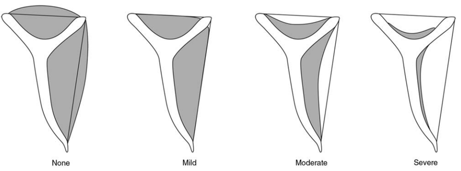
Illustration of the Warner method of evaluating rotator cuff atrophy based on T1-weighted sagittal oblique magnetic resonance images. The grade is determined by the amount of muscle above or below a line drawn from the edge of the coracoid to the tip of the scapular spine and a line from the superior aspect of the coracoid to the superior border of the scapular spine.
Hamada classification of rotator cuff arthropathy (53)
Acromiohumeral interval (AHI)
- Grade 1 – AHI over 6 mm
- Grade 2 – AHI < 5mm
- Grade 3 – Acetabulization
- Grade 4 – Acetabulization and narrowed GH joint
- Grade 5 - Acetabulization with humeral head collapse
Revision rotator cuff repair
The Sugaya classification for evaluation in revision rotator cuff repair is as follows:
- Type I - Sufficient thickness, homogeneous tendon (low signal on T2 images)
- Type II - Sufficient thickness, partial high-intensity from within the tendon
- Type III - Insufficient thickness without discontinuity
- Type IV - Minor discontinuity on more than one slice, suggesting a small tear
- Type V - Major discontinuity suggesting a moderate or large tear
On-Track/Off-Track Instability of the Shoulder (33,34,55,56)
Latarje or Remplissage procedures should be considered for anterior dislocations of the shoulder when there is evidence of an engaging 'off-track' Hill-Sachs lesion of the humerus, or if there is greater than 20% glenoid bone loss by X-ray, CT, or MRI.
The glenoid track, a zone of dynamic contact during arm elevation, is a unique biomechanical model that uses both glenoid and humeral head bone loss to predict subsequent risk of humeral head engagement and possible dislocation. An engaging Hill-Sachs bony defect, or ‘off-track’ lesion, is one in which the width of the bony defect is greater than the width of the glenoid track. Off-track engagement occurs when the medial margin of the Hill-Sachs defect engages the glenoid track. If there is bony loss of the glenoid as well, the glenoid track will proportionately be less, causing greater risk of engagement. A nonengaging, or ‘on-track’ Hill-Sachs lesion is one in which the width of the bony defect is less than the width of the glenoid track. Using preoperative CT or MR imaging, the glenoid track can identify individuals who are more likely to fail only a primary capsuloligamentous Bankart repair. Glenoid track evaluation shows that restoring the track (glenoid) to its normal width should be the first priority in restoring shoulder stability.
Subacromial Decompression (SAD)
There are 3 types of acromion anatomy according to Bigliani classification: type 1, flat (20%), type 2, curved (40%) and type 3, hooked, (40%). Acromioplasty involves removing bone from the undersurface of the acromion to change a type 3 (hooked) acromion to a type 1 (flat) acromion. Although debated for decades, current evidence concludes that there is no role for isolated acromioplasty (subacromial decompression), which prompted conversion of CPT code 29826 (acromioplasty, subacromial decompression) from an index, primary, "stand-alone" code to an "add-on" code only.
Summary of Evidence
Management of Rotator Cuff Injuries
- Study Design: This clinical practice guideline is based on a systematic review of current scientific and clinical research. The guideline contains 33 recommendations, including both diagnosis and treatment.
- Target Population: The guideline focuses on adults with rotator cuff injuries.
- Key Factors:
- Burden of Disease: Chronic shoulder pain affects approximately 8% of American adults, with rotator cuff pathology being the leading cause of shoulder-related disability.
- Etiology: Rotator cuff tears have two main causes: injury and degeneration.
- Incidence and Prevalence: Approximately 4.5 million patient visits related to shoulder pain occur each year in the United States.
- Management of Small to Medium Tears: Both physical therapy and surgical management result in notable improvement in patient-reported outcomes for patients with symptomatic small to medium full-thickness rotator cuff tears.
- Long-Term Nonsurgical Management: Patient-reported outcomes improve with physical therapy in symptomatic patients with full-thickness rotator cuff tears, but tear size, muscle atrophy, and fatty infiltration may progress over 5 to 10 years.
- Surgical Management: Healed rotator cuff repairs show improved patient-reported and functional outcomes compared to physical therapy and unhealed repairs.
- Acromioplasty and Rotator Cuff Repair: Routine use of acromioplasty as a concomitant treatment is not supported compared to arthroscopic repair alone for patients with small to medium-sized full-thickness rotator cuff tears.
- Diagnosis (Clinical Examination): Clinical examination can be useful to diagnose or stratify patients with rotator cuff tears, but a combination of tests will increase diagnostic accuracy.
- Diagnosis (Imaging): MRI, MRA, and ultrasonography are useful adjuncts to a clinical examination for identifying rotator cuff tears.
- Post-Op Mobilization Timing: Similar postoperative clinical and patient-reported outcomes for small to medium-sized full-thickness rotator cuff tears between early and delayed mobilization.
Physical Examination Tests of the Shoulder
- Study Design: This is a systematic review and meta-analysis of diagnostic test performance for physical examination tests (PETS) of the shoulder. The study adheres to the principles outlined in the Cochrane guidelines and the PRISMA statement.
- Target Population: The study includes data from 11 articles that met the inclusion criteria, focusing on patients with shoulder complaints.
- Key Factors:
- SLAP Lesions: The Compression-Rotation test obtained the highest diagnostic odds ratio (DOR) among single PETS for SLAP lesions.
- Rotator Cuff Tears: The Supraspinatus test obtained the highest DOR for diagnosing any full-thickness rotator cuff tear.
- Subacromial Impingement Syndrome: The Hawkins test obtained the highest DOR for diagnosing subacromial impingement syndrome.
- Overall Validity: The pooled DOR for PETS for SLAP lesions was 1.38, indicating statistical validity.
- Clinical Performance: No single PETS showed superior clinical test performance, but a combination of tests may increase diagnostic accuracy.
Management of Rotator Cuff Injuries Evidence-Based Clinical Practice Guideline
- Study Design: This clinical practice guideline (CPG) is based on a systematic review of published studies regarding the management of rotator cuff injuries. The review was conducted between October 2023 and June 2024, with the final search performed on June 7, 2024.
- Target Population: The guideline addresses the management of rotator cuff injuries in adults. It is not intended for pediatric patients.
- Key Factors:
- Management of Small to Medium Tears: Both physical therapy and operative treatment result in significant improvement in patient-reported outcomes for patients with symptomatic small to medium full-thickness rotator cuff tears.
- Long-Term Non-Operative Management: Patient-reported outcomes improve with physical therapy in symptomatic patients with full-thickness rotator cuff tears. However, the tear size, muscle atrophy, and fatty infiltration may progress over 5 to 10 years with non-operative management.
- Operative Management vs. Non-Operative Management: Healed rotator cuff repairs show improved patient-reported and functional outcomes compared to physical therapy and unhealed repairs.
- Acromioplasty & Rotator Cuff Repair: Routine use of acromioplasty as a concomitant treatment is not suggested for therapeutic benefit compared to arthroscopic repair alone for patients with small to medium-sized full-thickness rotator cuff tears.
- Diagnosis (Clinical Examination): Clinical examination can be useful to diagnose or stratify patients with rotator cuff tears; however, a combination of tests will increase diagnostic accuracy.
- Diagnosis (Imaging): MRI, MRA, CT, and ultrasound are useful adjuncts to a clinical exam and radiographs for identifying rotator cuff tears.
- Post-Op Mobilization Timing: Postoperative clinical and patient-reported outcomes are similar for small to medium-sized full-thickness rotator cuff tears managed with early or delayed mobilization.
Shared Findings:
- Effectiveness of Physical Therapy and Surgical Management:
- All three documents agree that both physical therapy and surgical management result in significant improvements in patient-reported outcomes (PROs) for patients with symptomatic small to medium full-thickness rotator cuff tears.
- Long-term non-operative management with physical therapy improves PROs, but the tear size, muscle atrophy, and fatty infiltration may progress over 5 to 10 years.
- All three documents agree that both physical therapy and surgical management result in significant improvements in patient-reported outcomes (PROs) for patients with symptomatic small to medium full-thickness rotator cuff tears.
- Diagnosis and Imaging:
- Clinical examination and imaging techniques such as MRI, MRA, CT, and ultrasound are useful adjuncts for diagnosing rotator cuff tears.
- A combination of physical examination tests (PETS) increases diagnostic accuracy compared to any single test.
- Clinical examination and imaging techniques such as MRI, MRA, CT, and ultrasound are useful adjuncts for diagnosing rotator cuff tears.
- Post-Operative Management:
- Post-operative clinical and patient-reported outcomes are similar for small to medium-sized full-thickness rotator cuff tears managed with early or delayed mobilization.
- Routine use of acromioplasty as a concomitant treatment is not suggested for therapeutic benefit compared to arthroscopic repair alone.
- Post-operative clinical and patient-reported outcomes are similar for small to medium-sized full-thickness rotator cuff tears managed with early or delayed mobilization.
Differing Findings:
- Specific Physical Examination Tests:
- The Gismervik et al. (2017) study provides a detailed analysis of various PETS, highlighting the diagnostic odds ratio (DOR) for different tests. For example, the Supraspinatus test obtained the highest DOR for diagnosing any full-thickness rotator cuff tear.
- The Gismervik et al. (2017) study provides a detailed analysis of various PETS, highlighting the diagnostic odds ratio (DOR) for different tests. For example, the Supraspinatus test obtained the highest DOR for diagnosing any full-thickness rotator cuff tear.
- Biological Augmentation and Other Treatments:
- The Weber et al. (2020) document discusses the use of biological augmentation with platelet-derived products, noting that while it does not improve PROs, limited evidence supports its use in decreasing retear rates.
- The Weber et al. (2020) document discusses the use of biological augmentation with platelet-derived products, noting that while it does not improve PROs, limited evidence supports its use in decreasing retear rates.
- Specific Recommendations and Strength of Evidence:
- The Weber et al. (2020) document provides a detailed breakdown of recommendations with varying strengths of evidence, including strong, moderate, limited, and consensus recommendations.
- The Weber et al. (2020) document provides a detailed breakdown of recommendations with varying strengths of evidence, including strong, moderate, limited, and consensus recommendations.
In summary, the evidence suggests that shoulder arthroscopy, combined with appropriate diagnostic and post-operative management strategies, is a valuable approach for treating rotator cuff injuries. The detailed analysis of PETS in the Gismervik et al. (2017) study and the comprehensive recommendations in the Weber et al. (2020) document provide additional insights that can guide clinical decision-making.
References
1. Kashanchi KI, Nazemi AK, Komatsu DE, Wang ED. Smoking as a risk factor for complications following arthroscopic rotator cuff repair. JSES Int. 2021;5(1):83-87. doi:10.1016/j.jseint.2020.10.002
2. Yau WP. Smokers Achieved Minimal Clinically Important Difference for Visual Analog Scale and American Shoulder and Elbow Surgeons Scores at a Lower Rate Than Nonsmokers Even When Repaired Supraspinatus Tendons Were Intact on Postoperative Magnetic Resonance Imaging. Arthrosc Sports Med Rehabil. 2024;6(2). doi:10.1016/j.asmr.
3. Akgün D, Maziak N, Plachel F, et al. Diagnostic Arthroscopy for Detection of Periprosthetic Infection in Painful Shoulder Arthroplasty. Arthroscopy: The Journal of Arthroscopic & Related Surgery. 2019;35(9):2571-2577. doi:10.1016/j.arthro.2019.03.058
4. Guild T, Kuhn G, Rivers M, Cheski R, Trenhaile S, Izquierdo R. The Role of Arthroscopy in Painful Shoulder Arthroplasty: Is Revision Always Necessary? Arthroscopy: The Journal of Arthroscopic & Related Surgery. 2020;36(6):1508-1514. doi:10.1016/j.arthro.2020.01.045
5. Zhang K, Crum RJ, Samuelsson K, Cadet E, Ayeni OR, de SA D. In-Office Needle Arthroscopy: A Systematic Review of Indications and Clinical Utility. Arthroscopy: The Journal of Arthroscopic & Related Surgery. 2019;35(9):2709-2721. doi:10.1016/j.arthro.2019.03.045
6. Thiagarajan A, Nagaraj R, Marathe K. Correlation Between Clinical Diagnosis, MRI, and Arthroscopy in Diagnosing Shoulder Pathology. Cureus. Published online December 24, 2021. doi:10.7759/cureus.20654
7. Weber S, Chahal J. Management of Rotator Cuff Injuries. Journal of the American Academy of Orthopaedic Surgeons. 2020;28(5):e193-e201. doi:10.5435/JAAOS-D-19-00463
8. Thangarajah T, Lo IK. Optimal Management of Partial Thickness Rotator Cuff Tears: Clinical Considerations and Practical Management. Orthop Res Rev. 2022;Volume 14:59-70. doi:10.2147/ORR.S348726
9. Gismervik S, Drogset JO, Granviken F, Rø M, Leivseth G. Physical examination tests of the shoulder: A systematic review and meta-analysis of diagnostic test performance. BMC Musculoskelet Disord. 2017;18(1). doi:10.1186/s12891-017-1400-0
10. Apostolopoulos AP, Angelis S, Yellapragada RK, et al. The Sensitivity of Magnetic Resonance Imaging and Ultrasonography in Detecting Rotator Cuff Tears. Cureus. Published online May 1, 2019. doi:10.7759/cureus.4581
11. Yubran AP, Pesquera LC, Juan ELS, et al. Rotator cuff tear patterns: MRI appearance and its surgical relevance. Insights Imaging. 2024;15(1). doi:10.1186/s13244-024-01607-w
12. Management of Rotator Cuff Injuries Evidence-Based Clinical Practice Guideline. American Academy of Orthopaedic Surgeons. Published online 2025. Accessed November 16, 2025. aaos.org/rccpg2025
13. Darbandi AD, Cohn M, Credille K, et al. A Systematic Review and Meta-analysis of Risk Factors for the Increased Incidence of Revision Surgery After Arthroscopic Rotator Cuff Repair. American Journal of Sports Medicine. 2024;52(5):1374-1383. doi:10.1177/03635465231182993
14. Traven SA, Brinton D, Simpson KN, et al. Preoperative Shoulder Injections Are Associated With Increased Risk of Revision Rotator Cuff Repair. Arthroscopy - Journal of Arthroscopic and Related Surgery. 2019;35(3):706-713. doi:10.1016/j.arthro.2018.10.107
15. Werner BC, Cancienne JM, Burrus MT, Griffin JW, Gwathmey FW, Brockmeier SF. The timing of elective shoulder surgery after shoulder injection affects postoperative infection risk in Medicare patients. J Shoulder Elbow Surg. 2016;25(3):390-397. doi:10.1016/j.jse.2015.08.039
16. Lucenti L, Panvini FMC, de Cristo C, et al. Do Preoperative Corticosteroid Injections Increase the Risk of Infection after Shoulder Arthroscopy or Shoulder Arthroplasty? A Systematic Review. Healthcare (Switzerland). 2024;12(5). doi:10.3390/healthcare12050543
17. Katepun S, Boonsun P, Boonsaeng WS, Apivatgaroon A. Reliability of the Single-Arm and Double-Arm Jobe Test for the Diagnosis of Full-Thickness Supraspinatus Tendon Tear. Orthop J Sports Med. 2023;11(8). doi:10.1177/23259671231187631
18. Kuzel BR, Grindel S, Papandrea R, Ziegler D. Fatty infiltration and rotator cuff atrophy. Journal of the American Academy of Orthopaedic Surgeons. 2013;21(10):613-623. doi:10.5435/JAAOS-21-10-613
19. Naimark M, Trinh T, Robbins C, et al. Effect of Muscle Quality on Operative and Nonoperative Treatment of Rotator Cuff Tears. Orthop J Sports Med. 2019;7(8). doi:10.1177/2325967119863010
20. Sheth MM, Shah AA. Massive and Irreparable Rotator Cuff Tears: A Review of Current Definitions and Concepts. Orthop J Sports Med. 2023;11(5). doi:10.1177/23259671231154452
21. Ghasemi SA, McCahon JAS, Yoo JC, et al. Subscapularis tear classification implications regarding treatment and outcomes: consensus decision-making. JSES Reviews, Reports, and Techniques. 2023;3(2):201-208. doi:10.1016/j.xrrt.2022.12.004
22. Mahatme RJ, Modrak M, Wilhelm C, et al. Glenohumeral Superior Translation and Subacromial Contract Pressure Are Both Improved With Superior Capsular Reconstruction: A Systematic Review and Meta-analysis of Biomechanical Investigations. Arthroscopy - Journal of Arthroscopic and Related Surgery. 2024;40(4):1279-1287. doi:10.1016/j.arthro.2023.08.025
23. Claro R, Fonte H. Superior capsular reconstruction: current evidence and limits. EFORT Open Rev. 2023;8(5):340-350. doi:10.1530/EOR-23-0027
24. Strauss EJ, McCormack RA, Onyekwelu I, Rokito AS. Management of failed arthroscopic rotator cuff repair. Journal of the American Academy of Orthopaedic Surgeons. 2012;20(5):301-309. doi:10.5435/JAAOS-20-05-301
25. Mandaleson A. Re-tears after rotator cuff repair: Current concepts review. J Clin Orthop Trauma. 2021;19:168-174. doi:10.1016/j.jcot.2021.05.019
26. Varacallo M, Tapscott DC, Mair SD. Superior Labrum Anterior Posterior Lesions. StatPearls. Published online August 4, 2023. https://www.ncbi.nlm.nih.gov/books/NBK538284/
27. Erickson J, Lavery K, Monica J, Gatt C, Dhawan A. Surgical treatment of symptomatic superior labrum anterior-posterior tears in patients older than 40 years: A systematic review. American Journal of Sports Medicine. 2015;43(5):1274-1282. doi:10.1177/0363546514536874
28. Dean RS, Onsen L, Lima J, Hutchinson MR. Physical Examination Maneuvers for SLAP Lesions: A Systematic Review and Meta-analysis of Individual and Combinations of Maneuvers. Am J Sports Med. 2023;51(11):3042-3052. doi:10.1177/03635465221100977
29. Familiari F, Huri G, Simonetta R, McFarland EG. SLAP lesions: Current controversies. EFORT Open Rev. 2019;4(1):25-32. doi:10.1302/2058-5241.4.180033
30. Hester WA, O’Brien MJ, Heard WMR, Savoie FH. Current Concepts in the Evaluation and Management of Type II Superior Labral Lesions of the Shoulder. Open Orthop J. 2018;12(1):331-341. doi:10.2174/1874325001812010331
31. Tupe RN, Tiwari V. Anteroinferior Glenoid Labrum Lesion (Bankart Lesion). StatPearls. Published online August 3, 2023. https://www.ncbi.nlm.nih.gov/books/NBK587359/
32. Pawłuś N, Kanak M, Frankiewicz A, et al. Remplissage May Decrease the Redislocation Rate After Arthroscopic Bankart Repair in Patients With an Engaging Hill-Sachs Defect: A Systematic Review and Meta-analysis of Studies With Minimal 2-Year Follow-up. American Journal of Sports Medicine. 2025;53(3):717-726. doi:10.1177/03635465241249492
33. Min KS, Horng J, Cruz C, Ahn HJ, Patzkowski J. Glenoid Bone Loss in Recurrent Shoulder Instability after Arthroscopic Bankart Repair: A Systematic Review. Journal of Bone and Joint Surgery. 2023;105(22):1815-1821. doi:10.2106/JBJS.23.00388
34. Momaya AM, Tokish JM. Applying the Glenoid Track Concept in the Management of Patients with Anterior Shoulder Instability. Curr Rev Musculoskelet Med. 2017;10(4):463-468. doi:10.1007/s12178-017-9436-1
35. Doehrmann R, Frush TJ, Frush TJ. Posterior Shoulder Instability. StatPearls. Published online July 10, 2023. https://www.ncbi.nlm.nih.gov/books/NBK557648/
36. Hurley ET, Aman ZS, Doyle TR, et al. Posterior Shoulder Instability, Part I—Diagnosis, Nonoperative Management, and Labral Repair for Posterior Shoulder Instability—An International Expert Delphi Consensus Statement. Arthroscopy - Journal of Arthroscopic and Related Surgery. 2025;41(2):166-180.e11. doi:10.1016/j.arthro.2024.04.035
37. Ireland ML, Hatzenbuehler JR. Superior labrum anterior to posterior (SLAP) tears. UpToDate. Published online May 11, 2023. https://www.uptodate.com/contents/superior-labrum-anterior-to-posterior-slap-tears
38. Johnson DJ, Tadi P. Multidirectional Shoulder Instability Continuing Education Activity.; 2023. Accessed November 9, 2025. https://www.ncbi.nlm.nih.gov/books/NBK557726/
39. Gerber C, Nyffeler RW. Classification of Glenohumeral Joint Instability. Clin Orthop Relat Res. 2002;400(400):65-76. doi:10.1097/00003086-200207000-00009
40. Pandey V, Madi S. Clinical Guidelines in the Management of Frozen Shoulder: An Update! Indian J Orthop. 2021;55(2):299-309. doi:10.1007/s
41. St Angelo JM, Taqi M, Fabiano SE. Adhesive Capsulitis. StatPearls. Published online August 4, 2023. https://www.ncbi.nlm.nih.gov/books/NBK532955/
42. Flores D V., Goes PK, Gómez CM, Umpire DF, Pathria MN. Imaging of the Acromioclavicular Joint: Anatomy, Function, Pathologic Features, and Treatment. RadioGraphics. 2020;40(5):1355-1382. doi:10.1148/rg.2020200039
43. Welch M, Rankin S, How Saw Keng M, Woods D. A systematic review of the treatment of primary acromioclavicular joint osteoarthritis. Shoulder Elbow. 2024;16(2):129-144. doi:10.1177/17585732231157090
44. Chen RE, Voloshin I. Long Head of Biceps Injury: Treatment Options and Decision Making. Sports Med Arthrosc Rev. 2018;26(3):139-144. doi:10.1097/JSA.0000000000000206
45. Hsu D, Anand P, Mabrouk A, Chang KV. Biceps Tendon Rupture. StatPearls. Published online July 15, 2023. https://www.ncbi.nlm.nih.gov/books/NBK513235/
46. Panico L, Roy T, Namdari S. Long Head of the Biceps Tendon Ruptures Biomechanics, Clinical Ramifications, and Management. JBJS Rev. 2021;9(10). doi:10.2106/JBJS.RVW.21.00092
47. Franceschetti E, Giovannetti de Sanctis E, Palumbo A, et al. The management of the long head of the biceps in rotator cuff repair: A comparative study of high vs. subpectoral tenodesis. J Sport Health Sci. 2023;12(5):613-618. doi:10.1016/j.jshs.2020.08.004
48. Ranieri R, Nabergoj M, Xu L, et al. Complications of Long Head of the Biceps Tenotomy in Association with Arthroscopic Rotator Cuff Repair: Risk Factors and Influence on Outcomes. J Clin Med. 2022;11(19). doi:10.3390/jcm11195657
49. Frank RM, Cotter EJ, Strauss EJ, Jazrawi LM, Romeo AA. Management of Biceps Tendon Pathology: From the Glenoid to the Radial Tuberosity. Journal of the American Academy of Orthopaedic Surgeons. 2018;26(4):e77-e89. doi:10.5435/JAAOS-D-17-00085
50. Habusta SF, Mabrouk A, Tuck JA. Synovial Chondromatosis. StatPearls. Published online April 22, 2023. https://www.ncbi.nlm.nih.gov/books/NBK470463/
51. Beard DJ, Rees JL, Cook JA, et al. Arthroscopic subacromial decompression for subacromial shoulder pain (CSAW): a multicentre, pragmatic, parallel group, placebo-controlled, three-group, randomised surgical trial. The Lancet. 2018;391(10118):329-338. doi:10.1016/S0140-6736(17)32457-1
52. Paavola M, Kanto K, Ranstam J, et al. Subacromial decompression versus diagnostic arthroscopy for shoulder impingement: A 5-year follow-up of a randomised, placebo surgery controlled clinical trial. Br J Sports Med. 2021;55(2):99-107. doi:10.1136/bjsports-2020-102216
53. Brolin TJ, Updegrove GF, Horneff JG. Classifications in Brief: Hamada Classification of Massive Rotator Cuff Tears. Clin Orthop Relat Res. 2017;475(11):2819-2823. doi:10.1007/s11999-017-5340-7
54. Sugaya H, Maeda K, Matsuki K, Moriishi J. Functional and Structural Outcome After Arthroscopic Full-Thickness Rotator Cuff Repair: Single-Row Versus Dual-Row Fixation. Arthroscopy. 2005;21(11):1307-1316. doi:10.1016/j.arthro.2005.08.011
55. Pécora JOR, Neves Junior AT, Roesler CRM, et al. Glenoid track evaluation by a validated finite-element shoulder numerical model. Orthopaedics and Traumatology: Surgery and Research. 2020;106(4):735-742. doi:10.1016/j.otsr.2020.03.004
56. Trivedi S, Pomerantz ML, Gross D, Golijanan P, Provencher MT. Shoulder instability in the setting of bipolar (glenoid and humeral head) bone loss: The glenoid track concept. In: Clinical Orthopaedics and Related Research. Vol 472. Springer New York LLC; 2014:2352-2362. doi:10.1007/s11999-014-3589-7
Coding Section
| CPT | Code | Description |
|
|
23120 |
Claviculectomy; partial |
|
|
23125 |
Claviculectomy; total |
|
|
23130 |
Acromioplasty or acromionectomy, partial, with or without coracoacromial ligament release |
|
|
23405 |
Tenotomy, shoulder area; single tendon |
|
|
23410 |
Repair of ruptured musculotendinous cuff (eg, rotator cuff) open; acute |
|
|
23412 |
Repair of ruptured musculotendinous cuff (eg, rotator cuff) open; chronic |
|
|
23415 |
Coracoacromial ligament release, with or without acromioplasty |
|
|
23420 |
Reconstruction of complete shoulder (rotator) cuff avulsion, chronic (includes acromioplasty) |
|
|
23430 |
Tenodesis of long tendon of biceps |
|
|
23450 |
Capsulorrhaphy, anterior; putti-platt procedure or magnuson |
|
|
23455 |
Capsulorrhaphy, anterior; with labral repair (eg, bankart procedure) |
|
|
23460 |
Capsulorrhaphy, anterior, any type; with bone block |
|
|
23462 |
Capsulorrhaphy, anterior, any type; with coracoid process transfer |
|
|
23465 |
Capsulorrhaphy, glenohumeral joint, posterior, with or without bone block |
|
|
23466 |
Capsulorrhaphy, glenohumeral joint, any type multi-directional instability |
|
|
23700 |
Manipulation under anesthesia, shoulder joint, including application of fixation apparatus (dislocation excluded) |
|
|
29805 |
Arthroscopy, shoulder, diagnostic, with or without synovial biopsy (separate procedure) |
|
|
29806 |
Arthroscopy, shoulder, surgical; capsulorrhaphy |
|
|
29807 |
Arthroscopy, shoulder, surgical; repair of slap lesion |
|
|
29819 |
Arthroscopy, shoulder, surgical; with removal of loose body or foreign body |
|
|
29820 |
Arthroscopy, shoulder, surgical; synovectomy, partial |
|
|
29821 |
Arthroscopy, shoulder, surgical; synovectomy, complete |
|
|
29822 |
Arthroscopy, shoulder, surgical; debridement, limited, 1 or 2 discrete structures (eg, humeral bone, humeral articular cartilage, glenoid bone, glenoid articular cartilage, biceps tendon, biceps anchor complex, labrum, articular capsule, articular side of the rotator cuff, bursal side of the rotator cuff, subacromial bursa, foreign body (ies)) |
|
|
29823 |
Arthroscopy, shoulder, surgical; debridement, extensive, 3 or more discrete structures (eg, humeral bone, humeral articular cartilage, glenoid bone, glenoid articular cartilage, biceps tendon, biceps anchor complex, labrum, articular capsule, articular side of the rotator cuff, bursal side of the rotator cuff, subacromial bursa, foreign body (ies)) |
|
|
29824 |
Arthroscopy, shoulder, surgical; distal claviculectomy including distal articular surface (mumford procedure) |
|
|
29825 |
Arthroscopy, shoulder, surgical; with lysis and resection of adhesions, with or without manipulation |
|
|
29826 |
Arthroscopy, shoulder, surgical; decompression of subacromial space with partial acromioplasty, with coracoacromial ligament (ie, arch) release, when performed (list separately in addition to code for primary procedure) |
|
|
29827 |
Arthroscopy, shoulder, surgical; with rotator cuff repair |
|
|
29828 |
Arthroscopy, shoulder, surgical; biceps tenodesis |
Procedure and diagnosis codes on Medical Policy documents are included only as a general reference tool for each policy. They may not be all-inclusive.
This medical policy was developed through consideration of peer-reviewed medical literature generally recognized by the relevant medical community, U.S. FDA approval status, nationally accepted standards of medical practice and accepted standards of medical practice in this community, and other nonaffiliated technology evaluation centers, reference to federal regulations, other plan medical policies, and accredited national guidelines.
"Current Procedural Terminology © American Medical Association. All Rights Reserved"
History From 2026 Forward
| 09/10/2026 | Annual review, updating policy for clarity and consistency. Updating definition of massive tear to > 5 and/or ≥ 2 tendons in rotator cuff repair, Hamada grad to ≤ 2 and the goutallier grad to ≥ 3 in RCR isolated superior capsular reconstruction, updated anterior inferior tears contraindications, and adding smoking cessation contraindication to RCT contraindications. Also updating general information, rationale, and references. |
| 04/01/2026 | New Policy |